What is Femoro-acetabular Impingement (FAI)?
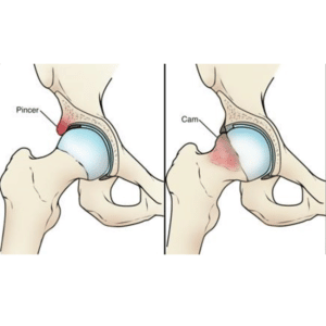
FAI occurs when the space between the ball (femur) and socket (acetabulum) in your hip joint is narrowed and the surrounding soft tissues become ‘pinched’.

FAI occurs when the space between the ball (femur) and socket (acetabulum) in your hip joint is narrowed and the surrounding soft tissues become ‘pinched’.
Pain can follow trauma but often arises with no clear cause and can progress with time. Pain is felt mostly into the groin, although can also be felt on the side of the pelvis. There can be a background ache felt but the greatest complaint is often a sharp or catching sensation in certain positions. More painful activities tend to be when flexing the hip or twisting the hip. Examples may include, although are not limited to:
There are four potential causes of Femoro-acetabular Impingement (FAI), often with more than one occurring at the same time:
In this scenario it is not the shape or structure of the joint causing the problem but in fact the way the joint is moving. The ball will sit towards the front of the socket and this reduces space on movement.
Surrounding the rim of the socket there is some thick, protective cartilage called fibro-cartilage which, amongst other things, deepens the socket. If there is a sudden onset of pain associated with a weight bearing twisting injury to the hip it is possible to damage the labrum.
The shape of the ball and socket joint is shown in the image above. In this scenario the femoral neck is thickened and so, on bringing the hip up, it comes into contact with the socket more rapidly.
This is opposite of a Cam impingement. Here there can be small bony projections called osteophytes from the edge of the socket bringing the rim of the socket closer to the ball.
The diagnostic process consists of three parts:
It is thought that often there is more than one factor contributing to FAI symptoms. Some people with Cam and pincer lesions do not experience pain. How the hip moves (biomechanics) is believed to play a role in most FAI cases. For this reason the first line of treatment for the majority of cases is physiotherapy.
Physiotherapy can be challenging initially and can even cause some pain. The aim of physiotherapy will be to correct muscle imbalance (strengthen weak muscles and stretch tight muscles and soft tissues). The main part of physiotherapy will be a home exercise programme.
Medication (such as simple painkillers or anti-inflammatories) may be useful. A pharmacist or your healthcare practitioner can help advise you what to take if needed. You can find further information here on what medications you could take here:
https://www.nhs.uk/medicines/paracetamol-for-adults/how-and-when-to-take-paracetamol-for-adults/
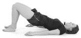
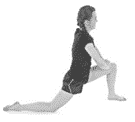
Exercise to manage FAI will depend on contributing factors and so will be tailored to the individual during physiotherapy sessions. Below are some exercises you can start in advance of your physiotherapy sessions.
If you have followed at least a three-month course of conservative management including physiotherapy, and have a clinical history and evidence on scans of FAI your healthcare practitioner may discuss other treatment options with you.
Joint injections have been shown to give symptom relief for up to six months in FAI.
Due to the nature of the hip joint they will need to be completed with fluoroscopic guidance so are undertaken in a radiology department. Since this is an invasive procedure there is some risk involved and may not be appropriate for everybody. Your healthcare practitioner will discuss this further with you as appropriate.
If conservative measures have failed to improve your pain there are surgical management options. If this is required your healthcare practitioner can discuss further with you.
Back to hip conditions